CLO 16: Describe the conditions that cause quantum mottle in a digital image

Too few x-ray photons (not enough mAs) = quantum noise

Image result for quantum noise x ray

What is quantum mottle? Grainy appearance on an image caused by underexposure

What does quantum mottle look like? Black and white speckles

How is quantum mottle caused? not enough x-ray photons

How do you get rid of quantum mottle? Increase technique (kVp & mAs)

CLO 7: Summarize the importance of proper positioning

Positioning is based on body part being imaged, condition of patient, and suspected disease. Body positioning is important because many of the internal structures superimpose over each other, therefore, certain positions are required to get a clear view of the area of concern.

Ex. Chest X-ray: The heart lies more anterior and a little to the left, this is why the PA projection is preformed; so the heart will be closer to the IR. A perpendicular CR is used at 72 SID. If any of these factors are altered we have the possibility of size distortion.

CLO 8: Discuss the impact of patient preparation on the resulting radiographic image

Ex. Abdomen

Patient needs to be fully changed in gown for an abdomen study. A pillow under their head and support under their knees for support will enhance patient comfort. A key factor in quality abdominal imaging is the prevention of motion; this is why breathing instructions are required for this exam. Using the shortest exposure time possible will help in this case. Before taking the exposure, ensure that the patient is following the breathing instructions and that you are allowing time for them to make all breathing movements. Abdominal radiographs are taken during the expiration portion, this will result in the diaphragm in a superior position for better visualization of abdominal structures.

Image result for inspiration vs expiration abdomen xray

CLO 6: Apply a process for evaluating images for acceptable limits of distortion, image artifacts, radiation fog, noise and gross exposure error

  1. Was the correct SID used?
  2. Does the projection demonstrate the least possible amount of size distortion?
  3. Does the projection demonstrate undesirable shape distortion?
  4. Was the part positioned with the least amount of OID possible?
  5. Does the projection demonstrate signs of undesirable patient motion or respiration?
  6. Are there anatomic artifacts demonstrated?
  7. Was scatter controlled with grids/collimation?
  8. Does the projection demonstrate quantum noise?
  9. Is there a decrease in contrast and detail visibility caused by fogging?
  10. Is the exposure indicator within the acceptable range for this system?
Image result for quantum noise x ray

CLO 4: Describe the role of the radiographer in image analysis.

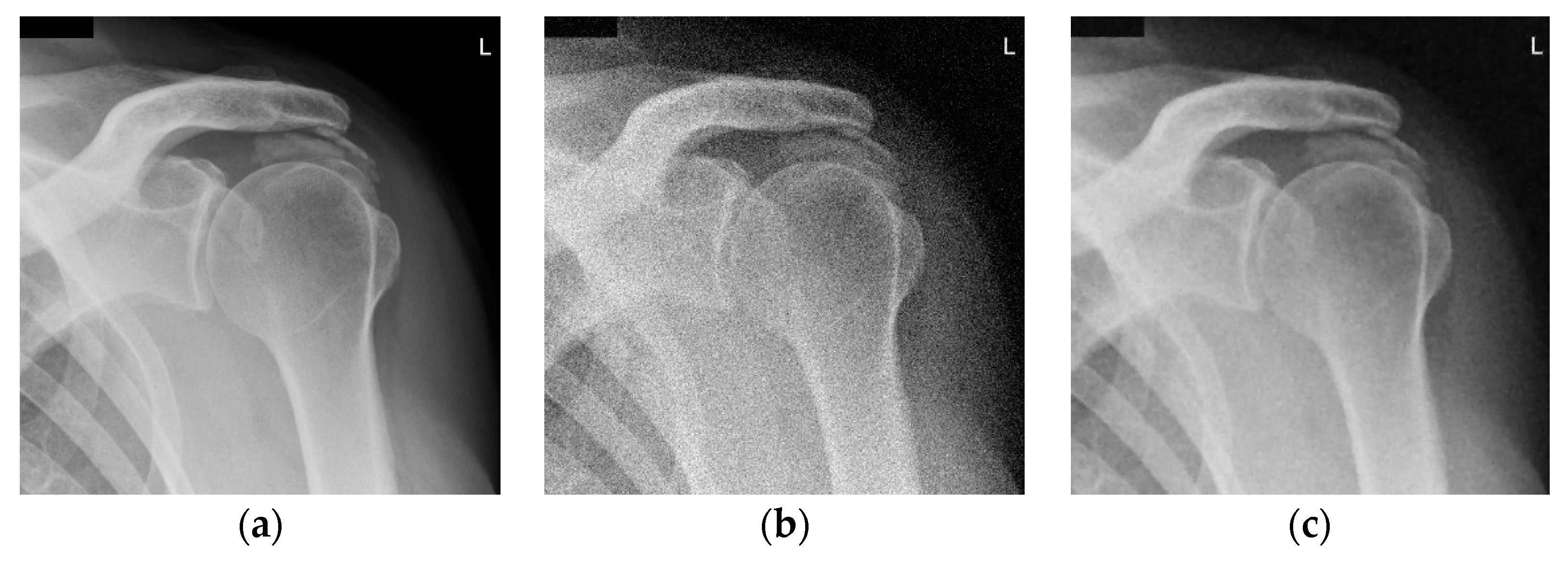
The technologist must be able to make an analysis of each radiographic image they take to determine if the image is acceptable. They must asses the following: Superimposition, adjacent structures, brightness, contrast, spatial resolution, magnification, and shape distortion. Although the technologist can not control involuntary motion such as a heartbeat, they can control voluntary motion such as discomfort by using sponges to keep their body in position to obtain the proper projection. When the technologist reviews their images they must be able to consider all normal appearances of the bodies structures.

Image result for poorly positioned PAhand

CLO 3: Describe an effective image analysis method


Evaluating a projection after the exposure is made:

  1. Demographic information is included
  2. Projection is accurately displayed
  3. Correct marker placement
  4. Appropriate collimation
  5. Relationships between the anatomic structures are accurate for the projection demonstrated
  6. Projection demonstrates maximum spatial resolution
  7. Radiation protection practice: Is gonadal shielding evident and accurately positioned when gonads are within the beam range?
  8. Is the exposure indicator within acceptable range for this system?
  9. Does this projection demonstrate quantum noise?
  10. Contrast resolution is optimal?
  11. No preventable artifacts are present

CLO 8: Adapt general procedural considerations to specific clinical settings.

“The technologist is expected to consider each patient’s general physical condition and clinical history. If the technologist feels the routines should be compromised, he/she should consult with a radiologist or technical supervisor.”

Body habitus: Hypersthenic, sthenic, hyposthenic, asthenic

4 quadrants of the abdomen: RUQ, RLQ, LUQ, LLQ

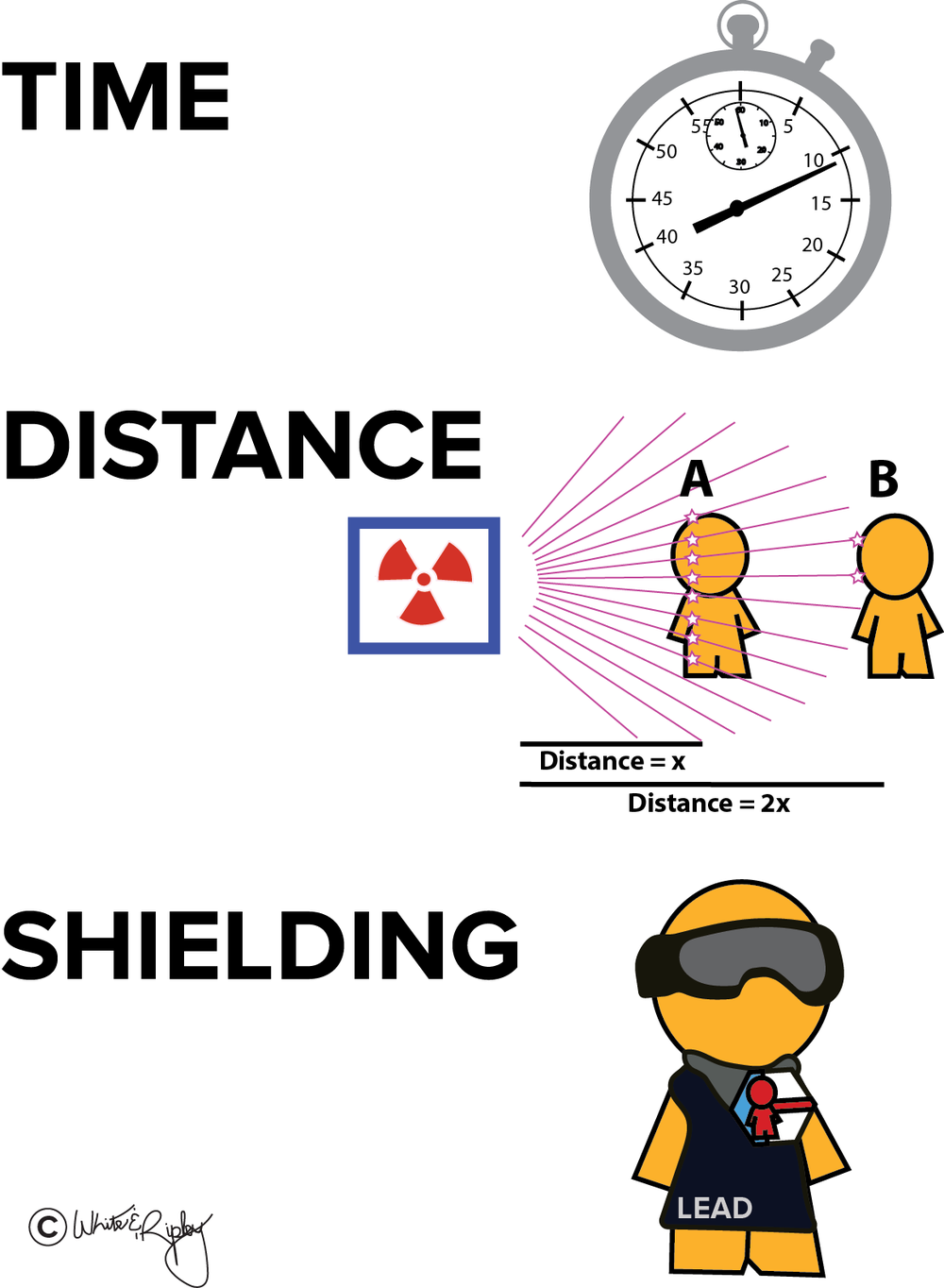
Angulation of CR may be required: to avoid superimposition of structures, to project through certain articulations, to avoid foreshortening

Portables: Alert all individuals in vicinity, before exposure is made

Isolation Case (Direct):

  • check with nurse for instructions and necessary precautions
  • machine must be wiped down with saniwipe after the study is completed
  • 2 technologist are necessary (imaging plate placed in plastic bag)
Related image

CLO 16: Apply general radiation safety and protection practices associated with radiographic and examinations

Minimize the radiation dose to the patient, to themselves, and to others:

As Low As Reasonably Achievable (ALARA) Principle

  1. Maintain a safe distance from source of ionizing radiation exposure
  2. Maximize the use of shielding from ionizing radiation exposure
  3. Limit the size of the x-ray exposure field to the area of interest
  4. Select a combination of kVp and mAs to produce a diagnostic image while minimizing patient radiation exposure
  5. Technologist wear a personnel monitoring device (i.e.: badge dosimeter 
  6. If technologist must assist in holding patient – they must wear a lead apron and, if possible, thyroid shield. 
Image result for radiation protection in medical imaging

CLO 15: Describe the general purpose of radiographic studies

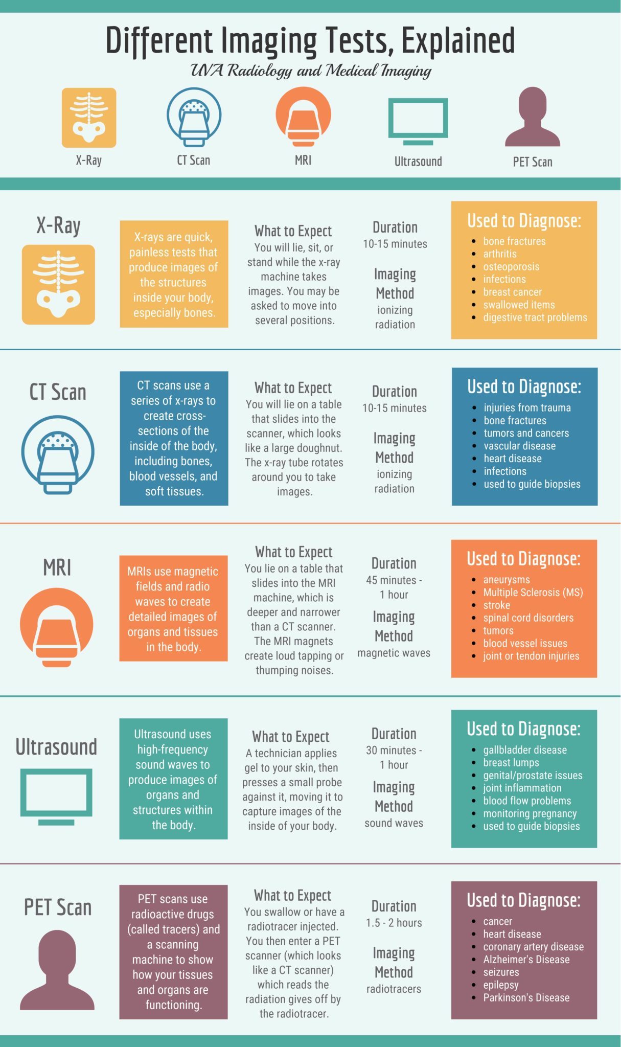
Radiographic studies are used to diagnose or treat patients by obtaining images of the internal structure of the body. These studies are used to examine an area where you’re experiencing pain/discomfort, monitor the progression of a diagnosed disease, and to follow up on a prescribed treatment. There are several types of diagnostic radiology exams.

Types of Imaging Exams, Explained
Design a site like this with WordPress.com
Get started