CLO 13: Discuss equipment and supplies necessary to complete basic radiographic and procedures

Why do radiographers follow a set of steps for each procedure🤔

☞To ensure patient safety, improve efficiency, and to reduce work errors.

Ex. Table or wall unit with an image receptor holder

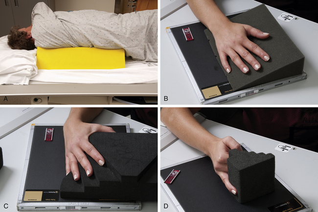
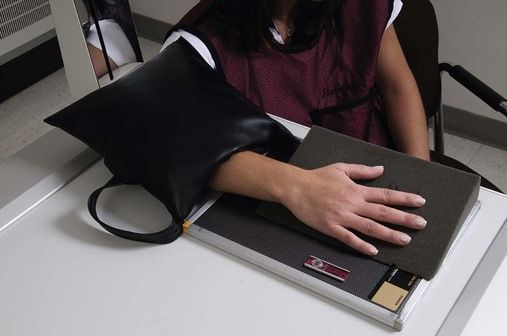
☑︎ Room preparation: clean equipment & accessories gathered such as grid, sponges, shield, etc.

☑︎ Choose IR: size & orientation

☑︎ Identify patient: name & DOB

☑︎ Obtain patient history

☑︎ Explain procedure

☑︎ Prepare patient: removing clothing and any artifact items

☑︎ Set Technical factors

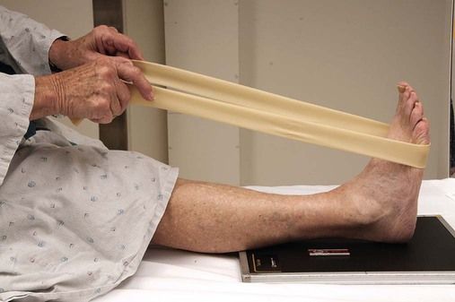
☑︎ Position patient: wedges/sponges may be used depending on position

☑︎ Set SID: varies depending on anatomy being imaged

☑︎ Align IR and CR

☑︎ Position part

☑︎ Collimate

☑︎ Correct marker placement

☑︎ Shield patient

☑︎ Provide any necessary patient instructions: breathing instructions

☑︎ Expose

☑︎ Evaluate radiograph

☑︎ Release patient: including exit instructions/follow-up information

A more complex procedure, such as fluoroscopy, will involve more equipment/supplies. For example, for a speech swallow, the technologist will gather controlled amounts of foods and liquids to prepare for this exam.

CLO 3: Discuss general procedural considerations for radiographic exams

✔︎Keep room clean to minimize transmission of infection

✔︎Use appropriate and required protection when there is a possibility of coming into contact with infectious organisms

✔︎Check with nursing staff or physician to obtain assistance and permission to move a part that has had surgery or is fractured

✔︎Verify correct procedure order

✔︎Observe patient conditions

✔︎If a patient has voluntary motion: use sponges to help support patient and ensure patient comfort

✔︎Shielding should be used when appropriate

✔︎Some examinations may require a grid 

CLO 1: Describe standard positioning terms

  • Anterior (ventral): forward or front part of body or organ
  • Posterior (Doral): back part of body or organ
  • Inferior: nearer the feet or situated below
  • superior: nearer the head or situated above
  • central: middle area or main part of an organ
  • lateral: parts away from median plane of body or away from the middle of another body part
  • medial: parts toward median plane of body or toward the middle of another body part
  • distal: parts farthest away from point of attachment
  • proximal: parts nearer point of attachment
  • external: parts outside an organ or on outside of body
  • internal: parts within or on the inside of an organ
  • dorm: the top or anterior surface of the foot or to the back or posterior surface of the hand
  • plantar: the sole of the foot
  • erect: either standing or sitting
  • decubitus: lying down
  • supine: lying on back
  • prone: lying face-down
  • lateral decubitus: lying on one
  • side right lateral: right side touches the cassette
  • left lateral: left side touches the cassette

CLO 4: Analyze the relationships of factors that control and affect image exposure.

To increase exposure to image receptor, increase mA, exposure time, or kVp. To decrease exposure to image receptor, decrease mA, exposure time, or kVp. Increasing the OID will decrease the exposure to the image receptor. Decreasing the OID will increase the exposure to the image receptor. A larger field size increases the amount of radiation reaching the image receptor. A smaller field size decreases the amount of radiation reaching the image receptor. 

To maintain exposure to image receptor:

Increase kVp 15% and decrease mAs by half

Decrease kVp 15% and increase mAs by two times

Ex. 60 kVp , 12.5 mAs

60 ⅹ .15 = 9 ➠ 60 -9 = 51 (New kVp)

12.5 ⅹ 2 = 25 (New mAs)

CLO 9: Differentiate between size and shape distortion

Size Distortion: increase in the object’s image size compared to its actual size

  • SID
  • OID

Increasing SID will decrease magnification 

Decreasing SID will increase magnification 

Increasing the OID increases magnification

Decreasing the OID decreases magnification

Shape Distortion: a misrepresentation of an object’s image shape

  • Central ray alignment of the x-ray tube, part, and image receptor 

CLO 8: Analyze the relationships of factors that control and affect spatial resolution

spatial resolution: refers to the smallest object that can be detected in a digital image

  • Increasing focal spot size – spatial resolution decreases
  • Decreasing focal spot size – spatial resolution increases
  • Increasing SID – increase spatial resolution
  • Decreasing SID – decrease spatial resolution
  • Increasing OID – decrease spatial resolution
  • Decreasing OID – increase spatial resolution

CLO 6: Analyze the relationship of factors that control and affect radiographic contrast

  • Increasing the OID – increase contrast 
  • Decreasing the OID – decrease contrast 
  • Grids – useful because this will have a less scatter or unwanted exposure on the image: increase the radiographic contrast. 
  • The decreased amount of scatter radiation reaching the image receptor results in higher radiographic contrast, *but it requires an increase in mAs. 
  • kVp controls radiographic contrast because it is the factor that determines the penetrating power of the x-ray beam. 

CLO 2: Discuss practical considerations in setting standards for acceptable image quality

  • IR/detector manufactures specify an expected range of x-rays exposure that will produce a good-quality image
  • A quality radiographic image accurately represents the anatomic area of interest
  • Brightness, contrast, spatial resolution, and distortion are all factors
  • X-ray tubes that have excessive or insufficient filtration may begin to affect image quality
A radiographic image must have sufficient brightness to visualize the anatomic structures of interest.
The radiograph must exhibit differences in the brightness levels or densities (image contrast) in order to differentiate among the anatomic tissues.
Spatial resolution refers to the smallest object that can be detected in a digital image. Shape distortion is a misrepresentation of an object’s image shape.

CLO 1: State the principles of x-ray production

The production of x-rays requires a rapidly moving stream of electrons that are suddenly decelerated or stopped. The cathode is heated, and the electrons are boiled off through the process of thermionic emission. There is a high voltage field between the cathode and anode. The electrons are then attracted to the anode, and move rapidly toward the positive end, where they are then decelerated or stopped.

Design a site like this with WordPress.com
Get started